File:Electron micrograph of neuromuscular junction (cross-section).jpg
此为最大尺寸。
Electron_micrograph_of_neuromuscular_junction_(cross-section).jpg (433 × 289像素,文件大小:95 KB,MIME类型:image/jpeg)
摘要
| 描述 |
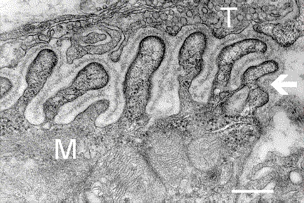
English: Electron micrograph showing a cross-section through the neuromuscular junction. T is the axon terminal, M is the muscle fiber. The arrow shows junctional folds with basal lamina. Postsynaptic densities are visible on the tips between the folds. The scale is 0.3 µm. |
| 日期 | Originally uploaded to en.wikipedia on 2006年3月10日. |
| 来源 | Synapse Web at the National Institute of Mental Health, National Institutes of Health; originally from en.wikipedia; description page is/was here. |
| 作者 | National Institute of Mental Health; originally uploaded by Nrets at en.wikipedia. |
许可协议
此圖像為美國衛生與公眾服務部下屬國家衛生院(NIH)僱員於公務中所拍攝或製作之作品。作為美國聯邦政府的作品,此圖像屬於公有領域。
 |
||
| 本文件已被确认为免除已知的著作权法限制(包括所有相关权利)。 | ||
原始上传日志
(All user names refer to en.wikipedia)
- 2006-03-10 20:07 Nrets 433×289×8 (97758 bytes) Electron micrograph showing a cross section through the neuromuscular junction. T is the axon terminal, M is the muscle fiber. The arrow shows junctional folds with basal lamina. Postsynaptic densities are visible on the tips between the folds. Scale is 0
文件历史
点击某个日期/时间查看对应时刻的文件。
| 日期/时间 | 缩略图 | 大小 | 用户 | 备注 | |
|---|---|---|---|---|---|
| 当前 | 2007年3月22日 (四) 03:41 | | 433 × 289(95 KB) | Fran Rogers | ((Information |Description=Electron micrograph showing a cross section through the neuromuscular junction. T is the axon terminal, M is the muscle fiber. The arrow shows junctional folds with basal lamina. Postsynaptic densities are visible on the tips be |
文件用途
以下页面使用本文件:
全域文件用途
以下其他wiki使用此文件:
- ar.wikipedia.org上的用途
- cs.wikipedia.org上的用途
- de.wikipedia.org上的用途
- es.wikipedia.org上的用途
- fa.wikipedia.org上的用途
- gl.wikipedia.org上的用途
- he.wikipedia.org上的用途
- ko.wikipedia.org上的用途
- pt.wikipedia.org上的用途
- ru.wikipedia.org上的用途
- uk.wikipedia.org上的用途
元数据
Text is available under the CC BY-SA 4.0 license; additional terms may apply.
Images, videos and audio are available under their respective licenses.
Cover photo is available under {{::mainImage.info.license.name || 'Unknown'}} license.
Cover photo is available under {{::mainImage.info.license.name || 'Unknown'}} license.
Credit:
(see original file).

.jpg)