蛋白质数据库
 | |
|---|---|
| 内容 | |
| 相关信息 | |
| 访问入口 | |
| 数据格式 | PDB |
| 网站 | |
| 工具 | |
| 其他 | |
蛋白质数据库(Protein Data Bank,简称PDB)是一个专门收录蛋白质及核酸的三维结构资料的数据库。由Worldwide Protein Data Bank监管。PDB可以经由网络免费访问,是结构生物学研究中的重要资源。为了确保PDB资料的完备与权威,各个主要的科学杂志、基金组织会要求科学家将自己的研究成果提交给PDB。在PDB的基础上,还发展出来若干依据不同原则对PDB结构数据进行分类的数据库,例如GO将PDB中的数据按基因进行了分类。这些资料和数据一般是世界各地的结构生物学家经由X射线晶体学或NMR光谱学实验所得,并释放到公有领域供公众免费使用。
历史
PDB的历史可以追溯到1971年,当时布鲁克黑文国家实验室的Walter Hamilton决定在Brookhaven建立这个数据库。1973年Hamilton去世后,Tom Koeztle接管了PDB。1994年1月,以色列魏茨曼科学研究学院的Joel Sussman被任命为PDB负责人。在1998年10月,PDB被移交给了Research Collaboratory for Structural Bioinformatics(RCSB),并与1999年6月移交完毕,新的负责人是罗格斯大学(Rutgers大学)(RCSB成员)的Helen M. Berman。2003年,PDB作为wwPDB的核心,成为了一个国际性组织。同时,wwPDB的其他成员,包括PDBe(欧洲)、RCSB(美国)、PDBj(日本)也为PDB提供了数据积累、处理和发布的中心。BMRB 于2006年加入[1]。值得一提的是,虽然PDB的数据是由世界各地的科学家提交的,但每条提交的数据都会经过wwPDB工作人员的审核与注解,并检验数据是否合理。PDB及其提供的软件现在对公众免费开放。
内容


PDB数据库每周更新(UTC+0 星期三)。 同样,PDB馆藏列表[2]也每周更新一次。 截至截至2017年3月14日[update],现有统计信息如下:
| 实验 方法 |
蛋白质 | 核酸 | 蛋白质/核酸 复合物 |
其他 | 总数 |
|---|---|---|---|---|---|
| X射线晶体学 | 106595 | 1820 | 5471 | 4 | 113890 |
| 核磁共振 | 10296 | 1190 | 241 | 8 | 11735 |
| 电子显微镜 | 1021 | 30 | 367 | 0 | 1418 |
| 混合 | 99 | 3 | 2 | 1 | 105 |
| 其他 | 181 | 4 | 6 | 13 | 204 |
| 总数: | 118192 | 3047 | 6087 | 26 | 127352 |
这些数据表明大多数结构是由X射线衍射确定的,但是现在约10%的结构由蛋白质NMR确定。 当使用X射线衍射时,获得蛋白质原子坐标的近似值,而通过NMR实验发现蛋白质原子对之间距离的估计值。 因此,在后一种情况下,通过求解距离几何问题来获得蛋白质的最终构象。 一些蛋白质由低温电子显微镜确定。 (单击原始表中的数字将显示由该方法确定的结构示例。)
上述结构因子文件的意义在于,对于具有结构文件的由X射线衍射确定的PDB结构,可以查看电子密度图。 这些结构的数据存储在“电子密度服务器”上[3][4] 。
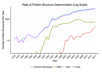
过去,PDB的结构数量以近似指数的速度增长,通过了1982年的100个注册结构里程碑,1993年的1,000个,1999年的10,000个,以及2014年的100,000个[5][6]。然而,自2007年以来,新蛋白质结构的积累速率似乎已经趋于稳定。
以下为PDB成员网站
参见
参考文献
- ^ BMRB - Biological Magnetic Resonance Bank. [2014-01-02]. (原始内容存档于2020-10-20).
- ^ PDB Current Holdings Breakdown. RCSB. [2018-04-22]. (原始内容存档于2007-07-04).
- ^ The Uppsala Electron Density Server. Uppsala University. [2013-04-06]. (原始内容存档于2021-03-21).
- ^ Kleywegt GJ, Harris MR, Zou JY, Taylor TC, Wählby A, Jones TA. The Uppsala Electron-Density Server. Acta Crystallogr D. Dec 2004, 60 (Pt 12 Pt 1): 2240–2249. PMID 15572777. doi:10.1107/S0907444904013253.
- ^ Anon. Hard data: It has been no small feat for the Protein Data Bank to stay relevant for 100,000 structures. Nature. 2014, 509 (7500): 260. PMID 24834514. doi:10.1038/509260a.
- ^ Content Growth Report. RCSB PDB. [2013-04-06]. (原始内容存档于2007-04-28).
外部链接
- (英文)PDB的官方网站(总站) (页面存档备份,存于互联网档案馆),局域网站(下)
- (英文)The Worldwide Protein Data Bank (wwPDB)— parent site to regional hosts (below)
- wwPDB Documentation (页面存档备份,存于互联网档案馆) — documentation on both the PDB and PDBML file formats
- (英文)Looking at Structures (页面存档备份,存于互联网档案馆) — The RCSB's introduction to crystallography
- (英文)PDBsum Home Page (页面存档备份,存于互联网档案馆) — Extracts data from other databases about PDB structures.
- (英文)Nucleic Acid Database, NDB (页面存档备份,存于互联网档案馆) — a PDB mirror especially for searching for nucleic acids
- (英文)Introductory PDB tutorial sponsored by PDB
- (英文)PDBe: Quick Tour on EBI Train OnLine (页面存档备份,存于互联网档案馆)
| |||||||||||||||||||||||||||||||||||||
Text is available under the CC BY-SA 4.0 license; additional terms may apply.
Images, videos and audio are available under their respective licenses.
