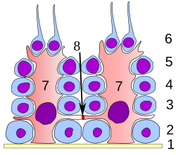
塞尔托利氏细胞

1: 基膜
2: 精原细胞
3: 初级精母细胞
4: 次级精母细胞
5: 精细胞
6: 成熟的精细胞
7: 塞尔托利氏细胞
8: 紧密连接

1: 细精管管腔
2: 精细胞
3: 精母细胞
4: 精原细胞
5: 塞尔托利氏细胞
6: 肌成纤维细胞
7: 莱氏细胞
8: 微血管
塞尔托利氏细胞(Sertoli cell),又名为塞托利细胞或史脱立细胞或塞透力细胞或支持细胞(sustentacular cell),是细精管一部分的睾丸的营养细胞。它是由促滤泡成熟激素(简称FSH)所启动,并在其细胞膜上有促滤泡成熟激素受体(FSHR)。
功能
塞尔托利氏细胞的主要功能是在精子发生过程中哺育成长中的精子细胞。因此,它亦被称为“母细胞”(不同于精母细胞)。它亦提供了分泌及结构性的支撑。
分泌
塞尔托利氏细胞会分泌以下的物质:
- 抗苗勒管激素(AMH)——于胎儿生命的早期就开始分泌。
- 抑制素及活化素——于青春期后分泌,配合调节促滤泡成熟激素(FSH)的分泌。
- 雄激素结合蛋白——与雄激素结合,维持生精小管内较高的雄激素水平,促进精子生成及精子成熟。
- 胶质源性神经营养因子(GDNF)——证实是助长精原细胞,以确保精细胞在产期时的自我更新。
- Ets相关分子——滋养在成人睾丸精原细胞内的精细胞。
- 转铁蛋白[1]
结构
塞尔托利氏细胞之间的连接形成了血睾屏障[2],血睾屏障是一个结构分隔睾丸空隙血液区及精细管内的向管腔区。塞尔托利氏细胞控制养份、激素及其他化合物进出睾丸的细管,且令向管腔区成为高度免疫的位点。
它亦负责确立及维持精原细胞的干细胞利基,以确保精子的更新及精原细胞在精子生成的过程中逐步分裂为成熟的生殖细胞,而最终为放出精子。
其他
在精子生成的成熟阶段,塞尔托利氏细胞会吞噬和消化精子生成过程中脱落的剩余胞质。[3]
睾丸支持细胞还可以将睾酮转化为雌二酮[3]
产生细胞
塞尔托利氏细胞的数目在青春期确定,成年男性不能产生新的塞尔托利氏细胞。[3]一些科学家最近发现在身体以外仍能培育这些细胞。[4][5]这提供了治疗男性不育缺憾的可能性。
命名
塞尔托利氏细胞是由发现这种细胞的意大利生理学家塞尔托利,他是在意大利帕维亚大学研究医药时发现的。[6]
他是于1862年在研究医药时用显微镜发现这种细胞。他于1865年发表这种细胞的描述,并以“像树的细胞”或“黏性细胞”来形容它。于1888年,其他科学家以他的名字来称呼这些细胞。
组织学
利用标准的染色法,是很容易将塞尔托利氏细胞与其他生殖上皮细胞混淆。而它最大的分别就是它那深色的细胞核。[7]

病理学
支持间质细胞瘤是属于卵巢瘤的性腺基质癌。
参见
参考
- ^ Xiong X, Wang A, Liu G, Liu H, Wang C, Xia T, Chen X, Yang K. Effects of p,p'-dichlorodiphenyldichloroethylene on the expressions of transferrin and androgen-binding protein in rat Sertoli cells.. Environ Res. 2006, 101 (3): 334–9. PMID 16380112.
- ^ de Kretser, David M.; Loveland, Kate; O’Bryan, Moira. Chapter 136 - Spermatogenesis. Jameson, J. Larry (编). Endocrinology: Adult and Pediatric (Seventh Edition). Philadelphia: W.B. Saunders. 2016-01-01: 2325–2353.e9. ISBN 978-0-323-18907-1. doi:10.1016/b978-0-323-18907-1.00136-0.
- ^ 3.0 3.1 3.2 Jones, Richard E.; Lopez, Kristin H. Chapter 4 - The Male Reproductive System. Jones, Richard E. (编). Human Reproductive Biology (Fourth Edition). San Diego: Academic Press. 2014-01-01: 67–83. ISBN 978-0-12-382184-3. doi:10.1016/b978-0-12-382184-3.00004-0.
- ^ Guo, Ying; Hai, Yanan; Yao, Chencheng; Chen, Zheng; Hou, Jingmei; Li, Zheng; He, Zuping. Long-term culture and significant expansion of human Sertoli cells whilst maintaining stable global phenotype and AKT and SMAD1/5 activation. Cell Communication and Signaling. 2015-03-25, 13 (1). ISSN 1478-811X. PMC 4380114
 . PMID 25880873. doi:10.1186/s12964-015-0101-2.
- ^ Lakpour, Mohammad Reza; Aghajanpour, Samaneh; Koruji, Morteza; Shahverdi, Abdolhossein; Sadighi-Gilani, Mohammad Ali; Sabbaghian, Marjan; Aflatoonian, Reza; Rajabian-Naghandar, Majid. Isolation, Culture and Characterization of Human Sertoli Cells by Flow Cytometry: Development of Procedure. Journal of Reproduction & Infertility. 2017, 18 (2). ISSN 2228-5482. PMC 5565911
 . PMID 28868245.
- ^ E. Sertoli: Dell’esistenza di particolari cellule ramificate nei canalicoli seminiferi del testicolo umano. Morgagni, 1865, 7: 31-40.
- ^ 存档副本. [2006-11-29]. (原始内容存档于2006-12-09).
外部链接
| |||||||||||||||||||||||||||||||||||||||||||||||||||||||||||||||||
| |||||||||||||||||||||||||||||||||||||||||||||||||||||||||
|
Text is available under the CC BY-SA 4.0 license; additional terms may apply.
Images, videos and audio are available under their respective licenses.
