For faster navigation, this Iframe is preloading the Wikiwand page for PD-L1.

PD-L1

CD274
PDBに登録されている構造
PDBオルソログ検索: RCSB PDBe PDBj
PDBのIDコード一覧

3BIK, 3BIS, 3FN3, 3SBW, 4Z18, 4ZQK, 5C3T, 5J89, 5J8O

識別子
記号CD274, B7-H, B7H1, PD-L1, PDCD1L1, PDCD1LG1, PDL1, CD274 molecule, Programmed cell death ligand 1, hPD-L1
外部IDOMIM: 605402 MGI: 1926446 HomoloGene: 8560 GeneCards: CD274
遺伝子の位置 (ヒト)
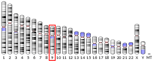
9番染色体 (ヒト)
染色体9番染色体 (ヒト)[1]
9番染色体 (ヒト)
CD274遺伝子の位置
CD274遺伝子の位置
バンドデータ無し開始点5,450,503 bp[1]
終点5,470,566 bp[1]
遺伝子の位置 (マウス)
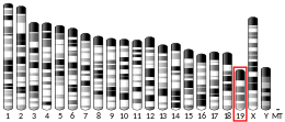
19番染色体 (マウス)
染色体19番染色体 (マウス)[2]
19番染色体 (マウス)
CD274遺伝子の位置
CD274遺伝子の位置
バンドデータ無し開始点29,344,855 bp[2]
終点29,365,495 bp[2]
遺伝子オントロジー
分子機能 血漿タンパク結合
細胞の構成要素 integral component of membrane

cell surface
エキソソーム
external side of plasma membrane
細胞内膜系
細胞膜
early endosome membrane
recycling endosome membrane
エンドソーム
生物学的プロセス negative regulation of interleukin-10 production
response to cytokine
negative regulation of interferon-gamma production
negative regulation of tumor necrosis factor superfamily cytokine production
positive regulation of cell migration
T cell costimulation
toxin transport
negative regulation of activated T cell proliferation
regulation of activated T cell proliferation
細胞表面受容体シグナル伝達経路
negative regulation of T cell proliferation
regulation of T cell apoptotic process
免疫応答
positive regulation of T cell proliferation
シグナル伝達
regulation of activated CD4-positive, alpha-beta T cell apoptotic process
positive regulation of activated CD8-positive, alpha-beta T cell apoptotic process
positive regulation of tolerance induction to tumor cell
negative regulation of CD8-positive, alpha-beta T cell activation
免疫系プロセス
negative regulation of CD4-positive, alpha-beta T cell proliferation
cellular response to lipopolysaccharide
adaptive immune response
出典:Amigo / QuickGO
オルソログ
ヒトマウス
Entrez
Ensembl
UniProt
RefSeq
(mRNA)

NM_001314029
NM_001267706
NM_014143

NM_021893

RefSeq
(タンパク質)

NP_001254635
NP_001300958
NP_054862

NP_068693

場所
(UCSC)
Chr 9: 5.45 – 5.47 MbChr 9: 29.34 – 29.37 Mb
PubMed検索[3][4]
ウィキデータ
閲覧/編集 ヒト閲覧/編集 マウス

PD-L1(プログラム細胞死リガンド1, Programmed death-ligand 1 別名:B7-H1)はPD-1リガンドである免疫チェックポイントタンパク質

概要

[編集]

PD-L1は発見当初はB7-H1として識別されていたが、後にPD-1リガンドである事が判明したのでPD-L1として改名された経緯がある。T細胞の活性を抑制もしくは停止させる(レセプター的な)免疫抑制機能を有するタンパク質分子であり、通常は抗原提示細胞の表面上に発現、また腫瘍細胞や腫瘍微小環境に存在する非形質転換細胞の細胞表面上においてもPD-L1が発現する事が知られており、同時に免疫チェックポイント阻害剤によるこれの遮断は腫瘍の増殖を阻害する事が知られ[5]免疫療法の結果を判断するバイオマーカーとしても注目される[6]

検出方法

[編集]

免疫染色によって検出する。

脚注

[編集]
  1. ^ a b c GRCh38: Ensembl release 89: ENSG00000120217 - Ensembl, May 2017
  2. ^ a b c GRCm38: Ensembl release 89: ENSMUSG00000016496 - Ensembl, May 2017
  3. ^ Human PubMed Reference:
  4. ^ Mouse PubMed Reference:
  5. ^ がん免疫療法と PD-1/PD-L1 チェックポイント・シグナル伝達”. abcam.co.jp. 2020年9月8日閲覧。
  6. ^ 癌治療効果のバイオマーカーとして有用な免疫チェックポイント分子 PD-L1”. abcam.co.jp. 2020年9月8日閲覧。

参考文献

[編集]
  • Mellati, Mahnaz, et al. "Anti–PD-L1 and anti–PDL-1 monoclonal antibodies causing type 1 diabetes." Diabetes care 38.9 (2015): e137-e138.
  • Rowe, Jared H., et al. "PDL-1 blockade impedes T cell expansion and protective immunity primed by attenuated Listeria monocytogenes." The Journal of Immunology 180.11 (2008): 7553-7557.
  • Bhadra, Rajarshi, et al. "Control of Toxoplasma reactivation by rescue of dysfunctional CD8+ T-cell response via PD-L1–PDL-1 blockade." Proceedings of the National Academy of Sciences 108.22 (2011): 9196-9201.
  • Yao, Zhi Q., et al. "T cell dysfunction by hepatitis C virus core protein involves PD-L1/PDL-1 signaling." Viral immunology 20.2 (2007): 276-287.
  • Sgambato, Assunta, et al. "Anti PD-L1 and PDL-1 immunotherapy in the treatment of advanced non-small cell lung cancer (NSCLC): a review on toxicity profile and its management." Current drug safety 11.1 (2016): 62-68.
  • Boasso, Adriano, et al. "PDL-1 upregulation on monocytes and T cells by HIV via type I interferon: restricted expression of type I interferon receptor by CCR5-expressing leukocytes." Clinical immunology 129.1 (2008): 132-144.
  • Sau, Samaresh, et al. "PDL-1 antibody drug conjugate for selective chemo-guided immune modulation of cancer." Cancers 11.2 (2019): 232.
  • Peng, YuFeng, Yvette Latchman, and Keith B. Elkon. "Ly6Clow monocytes differentiate into dendritic cells and cross-tolerize T cells through PDL-1." The Journal of Immunology 182.5 (2009): 2777-2785.
  • Channappanavar, Rudragouda, Brandon S. Twardy, and Susmit Suvas. "Blocking of PDL-1 interaction enhances primary and secondary CD8 T cell response to herpes simplex virus-1 infection." PloS one 7.7 (2012): e39757.
  • Wang, X. F., et al. "PD‐1/PDL 1 and CD 28/CD 80 pathways modulate natural killer T cell function to inhibit hepatitis B virus replication." Journal of Viral Hepatitis 20 (2013): 27-39.

関連項目

[編集]
{{bottomLinkPreText}} {{bottomLinkText}}
PD-L1
Listen to this article

This browser is not supported by Wikiwand :(
Wikiwand requires a browser with modern capabilities in order to provide you with the best reading experience.
Please download and use one of the following browsers:

This article was just edited, click to reload
This article has been deleted on Wikipedia (Why?)

Back to homepage

Please click Add in the dialog above
Please click Allow in the top-left corner,
then click Install Now in the dialog
Please click Open in the download dialog,
then click Install
Please click the "Downloads" icon in the Safari toolbar, open the first download in the list,
then click Install
{{::$root.activation.text}}

Install Wikiwand

Install on Chrome Install on Firefox
Don't forget to rate us

Tell your friends about Wikiwand!

Gmail Facebook Twitter Link

Enjoying Wikiwand?

Tell your friends and spread the love:
Share on Gmail Share on Facebook Share on Twitter Share on Buffer

Our magic isn't perfect

You can help our automatic cover photo selection by reporting an unsuitable photo.

This photo is visually disturbing This photo is not a good choice

Thank you for helping!


Your input will affect cover photo selection, along with input from other users.

X

Get ready for Wikiwand 2.0 🎉! the new version arrives on September 1st! Don't want to wait?