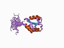
プロテインジスルフィドイソメラーゼ
| プロテインジスルフィドイソメラーゼ | |
|---|---|
 | |
| 識別子 | |
| 略号 | ? |
| InterPro | IPR005792 |
| Protein disulfide-isomerase | |||||||||
|---|---|---|---|---|---|---|---|---|---|
| 識別子 | |||||||||
| EC番号 | 5.3.4.1 | ||||||||
| CAS登録番号 | 37318-49-3 | ||||||||
| データベース | |||||||||
| IntEnz | IntEnz view | ||||||||
| BRENDA | BRENDA entry | ||||||||
| ExPASy | NiceZyme view | ||||||||
| KEGG | KEGG entry | ||||||||
| MetaCyc | metabolic pathway | ||||||||
| PRIAM | profile | ||||||||
| PDB構造 | RCSB PDB PDBj PDBe PDBsum | ||||||||
| 遺伝子オントロジー | AmiGO / QuickGO | ||||||||
| |||||||||
| protein disulfide isomerase family A, member 2 | |
|---|---|
| 識別子 | |
| 略号 | PDIA2 |
| 他の略号 | PDIP |
| Entrez | 64714 |
| HUGO | 14180 |
| OMIM | 608012 |
| RefSeq | NM_006849 |
| UniProt | Q13087 |
| 他のデータ | |
| 遺伝子座 | Chr. 16 p13.3 |
| protein disulfide isomerase family A, member 3 | |
|---|---|
| 識別子 | |
| 略号 | PDIA3 |
| 他の略号 | GRP58 |
| Entrez | 2923 |
| HUGO | 4606 |
| OMIM | 602046 |
| RefSeq | NM_005313 |
| UniProt | P30101 |
| 他のデータ | |
| 遺伝子座 | Chr. 15 q15 |
| protein disulfide isomerase family A, member 4 | |
|---|---|
| 識別子 | |
| 略号 | PDIA4 |
| Entrez | 9601 |
| HUGO | 30167 |
| RefSeq | NM_004911 |
| UniProt | P13667 |
| 他のデータ | |
| 遺伝子座 | Chr. 7 q35 |
| protein disulfide isomerase family A, member 5 | |
|---|---|
| 識別子 | |
| 略号 | PDIA5 |
| Entrez | 10954 |
| HUGO | 24811 |
| RefSeq | NM_006810 |
| UniProt | Q14554 |
| 他のデータ | |
| EC番号 (KEGG) | 5.3.4.1 |
| 遺伝子座 | Chr. 3 q21.1 |
| protein disulfide isomerase family A, member 6 | |
|---|---|
| 識別子 | |
| 略号 | PDIA6 |
| 他の略号 | TXNDC7 |
| Entrez | 10130 |
| HUGO | 30168 |
| RefSeq | NM_005742 |
| UniProt | Q15084 |
| 他のデータ | |
| 遺伝子座 | Chr. 2 p25.1 |
プロテインジスルフィドイソメラーゼまたはタンパク質ジスルフィドイソメラーゼ(英: protein disulfide isomerase、略称: PDI)は、真核生物の小胞体や細菌のペリプラズムに存在する酵素で、タンパク質がフォールディングする際にタンパク質内のシステイン残基の間のジスルフィド結合の形成と切断を触媒する[1][2][3]。PDIは、タンパク質が完全にフォールディングした状態で形成される適切なジスルフィド結合パターンの迅速な発見を可能にし、そのためフォールディングを触媒する酵素として作用する。
構造
[編集]PDIは2つのチオレドキシン様触媒ドメインと2つの非触媒ドメインからなり、各触媒ドメインには典型的なCGHCモチーフが存在する[4][5][6]。この構造はミトコンドリアの膜間腔で酸化的フォールディングを担う酵素の構造に類似している。こうした酵素の例としてはMia40があり、2つの触媒ドメインにはPDIのCGHCモチーフに類似したCX9Cモチーフが存在する[7]。細菌で酸化的フォールディングを担うDsbAもチオレドキシン様CXXCドメインを持っている[8]。

機能
[編集]タンパク質のフォールディング
[編集]PDIは酸化還元酵素と異性化酵素としての性質を持ち、どちらの活性もPDIに結合する基質の種類とPDIの酸化還元状態に依存している[4]。これらの活性はタンパク質の酸化的フォールディングを可能にする。酸化的フォールディングは新生タンパク質の還元型システイン残基の酸化を伴う過程であり、システイン残基が酸化されてジスルフィド結合が形成されることでタンパク質は安定化され、ネイティブ状態をとることができるようになる[4]。
通常の酸化的フォールディング機構と経路
[編集]
具体的には、PDIは小胞体でのタンパク質のフォールディングを担う[6]。フォールディングしていないタンパク質のシステイン残基はPDIの活性部位(CGHCモチーフ)内の2つのシステイン残基との間で混合型のジスルフィド結合を形成する。基質内で安定なジスルフィド結合が形成されると、PDIの活性部位の2つのシステイン残基は還元状態となって解離する[4]。その後、PDIはEro1(ER oxidoreductin 1)、VKOR(Vitamin K epoxide reductase)、グルタチオンペルオキシダーゼGpx7/8、ペルオキシレドキシンPrxIVといった再酸化タンパク質へ電子を伝達することで酸化型へと再生される[4][6][9][10]。PDIの主要な再酸化タンパク質はEro1であると考えられており、Ero1によるPDIの再酸化経路は他のタンパク質よりも理解が進んでいる[10]。Ero1はPDIから電子を受容し、その電子を小胞体の酸素分子へ供与する。その結果、過酸化水素が形成される[10]。
誤ったフォールディングを行ったタンパク質に対する活性
[編集]還元型のPDIは還元酵素活性または異性化酵素活性によって、基質に誤って導入されたジスルフィド結合の還元を触媒することができる[11]。還元酵素活性を利用する方法では、誤ったフォールディングを行った基質のジスルフィド結合は、グルタチオンやNADPHからの電子の移動によって還元型システイン残基へと変換される。その後、正しいシステイン残基の間で酸化的なジスルフィド結合の形成が行われ、正常なフォールディングが行われる。異性化酵素活性を利用する方法では、各活性部位のN末端近傍で基質分子内での官能基の再配置が行われる[4]。
酸化還元シグナル伝達
[編集]単細胞藻類であるクラミドモナスChlamydomonas reinhardtiiの葉緑体では、プロテインジスルフィドイソメラーゼRB60はpsbAの翻訳の光調節に関与するmRNA結合タンパク質複合体において、酸化還元センサーとして機能する。psbAは光化学系IIコアのD1タンパク質をコードする。プロテインジスルフィドイソメラーゼは葉緑体中で調節的なジスルフィド結合の形成にも関与している[12]。
他の機能
[編集]免疫系
[編集]PDIはMHCクラスI分子への抗原ペプチドのローディングを補助する。MHCクラスI分子は免疫応答において、抗原提示細胞によるペプチドの提示と関係している。
HIVがCD4陽性細胞へ感染する際、PDIはgp120タンパク質の結合の切断に関与していることが知られており、リンパ球や単球への感染に必要である[13]。
シャペロン活性
[編集]PDIの他の主要な機能としては、シャペロンとしての活性がある。C末端側の非触媒ドメイン(b'ドメイン)がミスフォールドタンパク質(誤ったフォールディングを行ったタンパク質)の結合とその後の分解を助ける[4]。この活性は3つの小胞体膜タンパク質、PERK(PKR-like endoplasmic reticulum kinase)、IRE1(inositol-requiring enzyme 1)、ATF6(activating transcription factor 6)によって調節される[4][14]。これらは小胞体中の高レベルのミスフォールドタンパク質に応答し、細胞内シグナル伝達経路を介してPDIのシャペロン活性を活性化する[4]。また、これらのシグナルはこうしたミスフォールドタンパク質の翻訳を不活性化する[4]。
活性のアッセイ
[編集]Insulin turbidity assay: PDIはインスリンの2本の鎖(a鎖とb鎖)の間の2つのジスルフィド結合を切断し、その結果b鎖は沈殿する。この沈殿は650 nmの波長でモニターすることができ、これによって間接的にPDIの活性をモニターすることができる[15]。このアッセイの感度はμMの範囲である。
ScRNase assay: PDIはさまざまな形でジスルフィド結合を形成した不活性なRNaseを、ネイティブ状態の活性のあるRNaseへ変換する[16]。このアッセイの感度はμMの範囲である。
Di-E-GSSG assay: このアッセイはpMの範囲でPDIを検出可能な蛍光定量アッセイであり、PDI活性を検出するアッセイの中で現在最も感度が高いものである[17]。Di-E-GSSGは酸化型グルタチオン(GSSG)に2つのエオシンが結合した分子であり、エオシンが近接して存在することでその蛍光は消光されるが、PDIによるジスルフィド結合の切断によって蛍光は70倍に増大する。
ストレスと阻害
[編集]ニトロソ化ストレスの影響
[編集]小胞体での酸化還元調節の異常はニトロソ化ストレスの増大をもたらす。神経細胞などの感受性細胞では、こうした細胞環境の変化によってチオールを含む酵素の機能喪失が引き起こされる[14]。より具体的には、PDIは活性部位のチオール基に一酸化窒素が付加されることでミスフォールドタンパク質を正しいフォールドへ修復することができなくなり、その結果、神経細胞にミスフォールドタンパク質が蓄積する。こうした現象はアルツハイマー病やパーキンソン病などの神経変性疾患の発症と関係している[4][14]。
阻害
[編集]PDIは多数の疾患に関与しているため、PDIに対する低分子阻害剤が開発されている。こうした分子はPDIの活性部位を不可逆的[18]または可逆的[19]に標的化する。
PDIの活性は赤ワインやグレープジュースによって阻害されることが示されており、フレンチパラドックスの説明となる可能性がある[20]。
メンバー
[編集]ヒトのプロテインジスルフィドイソメラーゼには次のようなものがある[3][21][22]。
- AGR2
- AGR3
- CASQ1
- CASQ2
- DNAJC10
- ERP27
- ERP29
- ERP44
- P4HB
- PDIA2
- PDIA3
- PDIA4
- PDIA5
- PDIA6
- PDIALT
- TMX1
- TMX2
- TMX3
- TMX4
- TXNDC5
- TXNDC12
出典
[編集]- ^ “Protein disulfide isomerase”. Biochimica et Biophysica Acta 1699 (1–2): 35–44. (June 2004). doi:10.1016/j.bbapap.2004.02.017. PMID 15158710.
- ^ “Protein disulfide isomerase: the structure of oxidative folding”. Trends in Biochemical Sciences 31 (8): 455–64. (August 2006). doi:10.1016/j.tibs.2006.06.001. PMID 16815710.
- ^ a b “The human protein disulfide isomerase gene family”. Human Genomics 6 (1): 6. (July 2012). doi:10.1186/1479-7364-6-6. PMC 3500226. PMID 23245351.
- ^ a b c d e f g h i j k “The Unfolded Protein Response and the Role of Protein Disulfide Isomerase in Neurodegeneration” (英語). Frontiers in Cell and Developmental Biology 3: 80. (2016). doi:10.3389/fcell.2015.00080. PMC 4705227. PMID 26779479.
- ^ “From structure to redox: The diverse functional roles of disulfides and implications in disease”. Proteomics 17 (6): n/a. (March 2017). doi:10.1002/pmic.201600391. PMC 5367942. PMID 28044432.
- ^ a b c “Protein disulfide isomerases: Redox connections in and out of the endoplasmic reticulum”. Archives of Biochemistry and Biophysics. The Chemistry of Redox Signaling 617: 106–119. (March 2017). doi:10.1016/j.abb.2016.11.007. PMID 27889386.
- ^ “Mitochondrial disulfide relay and its substrates: mechanisms in health and disease”. Cell and Tissue Research 367 (1): 59–72. (January 2017). doi:10.1007/s00441-016-2481-z. PMID 27543052.
- ^ “Structure of TcpG, the DsbA protein folding catalyst from Vibrio cholerae”. Journal of Molecular Biology 268 (1): 137–46. (April 1997). doi:10.1006/jmbi.1997.0940. PMID 9149147.
- ^ “Oxidative protein biogenesis and redox regulation in the mitochondrial intermembrane space”. Cell and Tissue Research 367 (1): 43–57. (January 2017). doi:10.1007/s00441-016-2488-5. PMC 5203823. PMID 27632163.
- ^ a b c “Thiol-disulfide exchange between the PDI family of oxidoreductases negates the requirement for an oxidase or reductase for each enzyme”. The Biochemical Journal 469 (2): 279–88. (July 2015). doi:10.1042/bj20141423. PMC 4613490. PMID 25989104.
- ^ “Substrate recognition by the protein disulfide isomerases”. The FEBS Journal 274 (20): 5223–34. (October 2007). doi:10.1111/j.1742-4658.2007.06058.x. PMID 17892489.
- ^ “Disulfide bond formation in chloroplasts”. Plant Science 175 (4): 459–466. (2008). doi:10.1016/j.plantsci.2008.05.011.
- ^ “Progress in targeting HIV-1 entry”. Drug Discovery Today 10 (16): 1085–94. (August 2005). doi:10.1016/S1359-6446(05)03550-6. PMID 16182193.
- ^ a b c “Redox-based therapeutics in neurodegenerative disease”. British Journal of Pharmacology 174 (12): 1750–1770. (June 2017). doi:10.1111/bph.13551. PMC 5446580. PMID 27477685.
- ^ “Protein disulfide-isomerase is a substrate for thioredoxin reductase and has thioredoxin-like activity”. The Journal of Biological Chemistry 265 (16): 9114–20. (June 1990). doi:10.1016/S0021-9258(19)38819-2. PMID 2188973.
- ^ “Catalysis of the oxidative folding of ribonuclease A by protein disulfide isomerase: dependence of the rate on the composition of the redox buffer”. Biochemistry 30 (3): 613–9. (January 1991). doi:10.1021/bi00217a004. PMID 1988050.
- ^ “Characterization of redox state and reductase activity of protein disulfide isomerase under different redox environments using a sensitive fluorescent assay”. Free Radical Biology & Medicine 43 (1): 62–70. (July 2007). doi:10.1016/j.freeradbiomed.2007.03.025. PMID 17561094.
- ^ “Inhibitors of protein disulfide isomerase suppress apoptosis induced by misfolded proteins”. Nature Chemical Biology 6 (12): 900–6. (December 2010). doi:10.1038/nchembio.467. PMC 3018711. PMID 21079601.
- ^ “Small molecule-induced oxidation of protein disulfide isomerase is neuroprotective”. Proceedings of the National Academy of Sciences of the United States of America 112 (17): E2245-52. (April 2015). Bibcode: 2015PNAS..112E2245K. doi:10.1073/pnas.1500439112. PMC 4418888. PMID 25848045.
- ^ “Revisiting the mechanistic basis of the French Paradox: Red wine inhibits the activity of protein disulfide isomerase in vitro”. Thrombosis Research 137: 169–173. (January 2016). doi:10.1016/j.thromres.2015.11.003. PMC 4706467. PMID 26585763.
- ^ “The human protein disulphide isomerase family: substrate interactions and functional properties”. EMBO Reports 6 (1): 28–32. (January 2005). doi:10.1038/sj.embor.7400311. PMC 1299221. PMID 15643448.
- ^ “The human PDI family: versatility packed into a single fold”. Biochimica et Biophysica Acta (BBA) - Molecular Cell Research 1783 (4): 535–48. (April 2008). doi:10.1016/j.bbamcr.2007.11.010. PMID 18093543.
外部リンク
[編集]Text is available under the CC BY-SA 4.0 license; additional terms may apply.
Images, videos and audio are available under their respective licenses.
