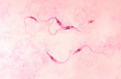
ギムザ染色

ギムザ染色(ギムザせんしょく、英: Giemsa stain)は、血液標本染色法の1つ。マラリア研究の先駆である医学者、グスタフ・フォン・ギムザ(Gustav von Giemsa、1867年 - 1948年)の名を取って「ギムザ染色」と呼ぶ。[1][2]
ドイツ・ハンブルクの熱帯病研究所にて、マラリア原虫の染色法として開発された。現在も臨床現場で広く用いられている。
利用
[編集]DNAのリン酸基に特異的であり、アデニン-チミン結合が多いDNA領域に結合する。ギムザ染色は染色体を染色するためにギムザ染色法(染色体地図)で用いられる。転座や再配列などの染色体異常を同定できる。
栄養型Trichomonas vaginalisを染色し、緑色の分泌物と湿潤環境下での運動性細胞を示す。
ギムザ染色は、ライト染色と組み合わせてライト-ギムザ染色を形成する場合などの差次染色でもある。ヒト細胞への病原性細菌の付着を研究するために使用できる。ヒトと細菌の細胞をそれぞれ紫とピンクに染める。マラリア[3]や他のスピロヘータや原虫の血液寄生虫の病理組織学的診断に用いることができる。キイロショウジョウバエのWolbachia細胞染色にも用いられる。
ギムザ染色は、末梢血塗抹標本および骨髄標本に対する古典的な血液フィルム染色法である。赤血球はピンク色、血小板は淡薄桃色、リンパ球細胞質は空色、単球細胞質は淡青色、白血球核クロマチンはマゼンタに染まる。また、ペスト菌の古典的な「安全ピン」の形を視覚化するのにも用いられる。
ギムザ染色は染色体の視覚化にも用いられる。これは、典型的な所見が「フクロウの目(owl-eye)」ウイルス封入体であるサイトメガロウイルス感染の検出に特に関連する[4]。
ギムザは真菌のヒストプラズマやクラミジアを染色し、肥満細胞の同定に用いることができる[5]。
手法
[編集]溶液はメチレンブルー、エオシンおよびアズールBの混合物であり、染色は通常市販のギムザ粉末から調製される。
顕微鏡スライド上の試料の薄膜を純メタノールに浸漬するか、あるいはスライド上に数滴メタノールを滴下して30秒間固定する。スライドを新しく調製した5%ギムザ染色液に20 - 30分間浸漬し(緊急時には10%溶液で5 - 10分使用可能)、次に水道水でフラッシュし、乾燥させる[6]。
脚注
[編集]- ^ Zipfel, E.; Grezes, J. -R.; Naujok, A.; Seiffert, W.; Wittekind, D. H.; Zimmermann, H. W. (1984). “Über Romanowsky-Farbstoffe und den Romanowsky-Giemsa-Effekt”. Histochemistry 81 (4): 337-351. doi:10.1007/bf00514328. ISSN 0301-5564.
- ^ Giemsa G (1904 Eine Vereinfachung und Vervollkommnung meiner Methylenblau-Eosin-Färbemethode zur Erzielung der Romanowsky-Nocht’schen Chromatinfärbung. Centralblatt für Bakteriologie I Abteilung 32, 307-313.
- ^ Shapiro HM, Mandy F (September 2007). "Cytometry in malaria: moving beyond Giemsa". Cytometry Part A. 71 (9): 643-5. doi:10.1002/cyto.a.20453, PMID 17712779.
- ^ Woods, G. L.; Walker, D. H. (July 1996). "Detection of infection or infectious agents by use of cytologic and histologic stains". Clinical Microbiology Reviews. 9 (3): 382-404. doi:10.1128/cmr.9.3.382, ISSN 0893-8512. PMC 172900. PMID 8809467.
- ^ Damsgaard TE, Olesen AB, Sørensen FB, Thestrup-Pedersen K, Schiøtz PO (April 1997). "Mast cells and atopic dermatitis. Stereological quantification of mast cells in atopic dermatitis and normal human skin". Arch. Dermatol. Res. 289 (5): 256-60. doi:10.1007/s004030050189, PMID 9164634.
- ^ "4.2.2.2. Giemsa stain". impact-malaria.com. Archived from the original on 2013-10-29. Retrieved 28 Oct 2013.
関連項目
[編集]Text is available under the CC BY-SA 4.0 license; additional terms may apply.
Images, videos and audio are available under their respective licenses.
