For faster navigation, this Iframe is preloading the Wikiwand page for चित्र:S cerevisiae under DIC microscopy.jpg.

चित्र:S cerevisiae under DIC microscopy.jpg
मूल चित्र((1,560 × 1,560 पिक्सेल, फ़ाइल का आकार: 610 KB, MIME प्रकार: image/jpeg))

सारांश

विवरण
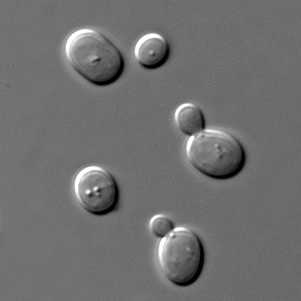
English: Saccharomyces cerevisiae cells in DIC microscopy. Imaging was performed with the Olympus BX61 microscope and a UPlanSApo 100× NA 1.40 oil immersion objective (Olympus). Pictures were acquired at room temperature in synthetic complete medium with a camera (SPOT; Diagnostic Instruments, Inc.) using MetaMorph software (MDS Analytical Technologies).
Polski: Komórki drożdży piekarniczych (Sacharomyces cerevisiae) widziane w różnicowej mikroskopii interferencyjnej. Zdjęcie wykonane w syntetycznej pożywce kompletnej za pomocą mikroskopu Olympus BX61 wyposażonego w obiektyw imersyjny (olej) 100x NA 1,40 (Olympus) sprzężonego z kamerą SPOT (Diagnostic Instruments, Inc.). Użyto programu MetaMorph software (MDS Analytical Technologies).
दिनांक
स्रोत अपना कार्य
लेखक Masur
अनुमति
(इस चित्र का पुनः उपयोग करना)
Public domain मैं, इस कार्य का/की कॉपीराइट धारक, इस कार्य को सार्वजनिक डोमेन में प्रकाशित करता/करती हूँ। यह पूरे विश्व में लागू होता है।
कुछ देशों में यह कानूनी तौर पर नहीं हो सकता है; ऐसा हो तो:
मैं सभी को इस कार्य का इस्तेमाल किसी भी उद्देश्य से, बिना किसी बाधाओं के इन शर्तों के कानून द्वारा अनिवार्य किए तक करने की अनुमति देता/देती हूँ।

Captions

Add a one-line explanation of what this file represents
Yeast of the species Saccharomyces cerevisiae

Items portrayed in this file

चित्रण

source of file अंग्रेज़ी

original creation by uploader अंग्रेज़ी

media type अंग्रेज़ी

image/jpeg

fabrication method अंग्रेज़ी

चित्र का इतिहास

फ़ाइलका पुराना अवतरण देखने के लिये दिनांक/समय पर क्लिक करें।

दिनांक/समयथंबनेलआकारसदस्यप्रतिक्रिया
वर्तमान21:06, 3 मार्च 201921:06, 3 मार्च 2019 के संस्करण का थंबनेल संस्करण1,560 × 1,560 (610 KB)Andrew Pertsevturn for the usual lighting
14:02, 14 जनवरी 201014:02, 14 जनवरी 2010 के संस्करण का थंबनेल संस्करण1,567 × 1,567 (540 KB)MasurI know that I shouldn't but the previous version has got such a bad quality that I cannot look at it, so I made a new one - the same yeast, the same microscope, more skills :)
08:30, 19 अगस्त 200608:30, 19 अगस्त 2006 के संस्करण का थंबनेल संस्करण500 × 500 (160 KB)MasurSacharomyces cerevisiae cells under DIC microscopy. Komórki drożdży piekarniczych widziane w różnicowej mikroskopii interferencyjnej.

चित्र का उपयोग

निम्नलिखित पन्ने इस चित्र से जुडते हैं :

चित्र का वैश्विक उपयोग

इस चित्र का उपयोग इन दूसरे विकियों में किया जाता है:

इस चित्र के वैश्विक उपयोग की अधिक जानकारी देखें।

मेटाडेटा

{{bottomLinkPreText}} {{bottomLinkText}}
चित्र:S cerevisiae under DIC microscopy.jpg
Listen to this article

This browser is not supported by Wikiwand :(
Wikiwand requires a browser with modern capabilities in order to provide you with the best reading experience.
Please download and use one of the following browsers:

This article was just edited, click to reload
This article has been deleted on Wikipedia (Why?)

Back to homepage

Please click Add in the dialog above
Please click Allow in the top-left corner,
then click Install Now in the dialog
Please click Open in the download dialog,
then click Install
Please click the "Downloads" icon in the Safari toolbar, open the first download in the list,
then click Install
{{::$root.activation.text}}

Install Wikiwand

Install on Chrome Install on Firefox
Don't forget to rate us

Tell your friends about Wikiwand!

Gmail Facebook Twitter Link

Enjoying Wikiwand?

Tell your friends and spread the love:
Share on Gmail Share on Facebook Share on Twitter Share on Buffer

Our magic isn't perfect

You can help our automatic cover photo selection by reporting an unsuitable photo.

This photo is visually disturbing This photo is not a good choice

Thank you for helping!


Your input will affect cover photo selection, along with input from other users.

X

Get ready for Wikiwand 2.0 🎉! the new version arrives on September 1st! Don't want to wait?