Lámina terminal
| Lámina terminal | ||
|---|---|---|
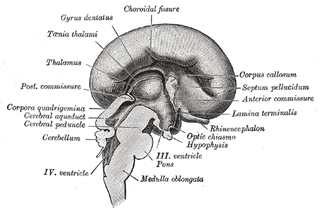
 Sección sagital mediana de cerebro de embrión humano de tres meses. (Lamina terminalis marcada en el centro a la izquierda). | ||
 Sección sagital mediana de cerebro de embrión humano de cuatro meses. (Lamina terminalis señalada en el centro a la derecha). | ||
| Nombre y clasificación | ||
| Latín | Lamina terminalis | |
| TA | A14.1.08.419 | |
|
| ||
La porción media de la pared del cerebro anterior está formada por una lámina delgada, la lámina terminal, que se extiende desde el agujero interventricular (agujero de Monro) hasta la escotadura de la base del tallo óptico (nervio óptico) y contiene el órgano vascular de la lámina terminal, que regula la concentración osmótica de la sangre. La lámina terminal es inmediatamente anterior al tubérculo cinéreo; juntos forman el tallo hipofisario.
La lámina terminal puede abrirse mediante neurocirugía endoscópica en un intento de crear una vía por la que pueda fluir el líquido cefalorraquídeo cuando una persona tiene hidrocefalia y cuando no es posible realizar una tercera ventriculostomía endoscópica, pero la eficacia de esta técnica no es segura.[1][2]
Se trata del extremo rostral (punta) del tubo neural (sistema nervioso central embriológico) en las primeras semanas de desarrollo. Si la lámina terminal no se cierra correctamente en esta fase del desarrollo, se producirá anencefalia.[3]
Imágenes adicionales
[editar]-
Aspecto mesal de un cerebro seccionado en el plano sagital medio.
-
Cerebro. Vista inferior.Disección profunda
Véase también
[editar]Referencias
[editar]- Este artículo incorpora texto de dominio público de la página 816 de la 20ª edición de Gray's Anatomy (1918)
- ↑ Oertel, J. M.; Vulcu, S; Schroeder, H. W.; Konerding, M. A.; Wagner, W; Gaab, M. R. (2010). «Endoscopic transventricular third ventriculostomy through the lamina terminalis». Journal of Neurosurgery 113 (6): 1261-9. PMID 20707616. doi:10.3171/2010.6.JNS09491.
- ↑ Komotar, R. J.; Hahn, D. K.; Kim, G. H.; Starke, R. M.; Garrett, M. C.; Merkow, M. B.; Otten, M. L.; Sciacca, R. R. et al. (2009). «Efficacy of lamina terminalis fenestration in reducing shunt-dependent hydrocephalus following aneurysmal subarachnoid hemorrhage: A systematic review. Clinical article». Journal of Neurosurgery 111 (1): 147-54. PMID 19284236. doi:10.3171/2009.1.JNS0821.
- ↑ Carlson, Neil R.; Birkett, Melissa A. (2017). Physiology of Behavior (12 edición). Ingestive Behavior: Pearson. p. 370. ISBN 9780134320823.
Enlaces externos
[editar]- Esta obra contiene una traducción derivada de «Lamina terminalis» de Wikipedia en inglés, concretamente de esta versión, publicada por sus editores bajo la Licencia de documentación libre de GNU y la Licencia Creative Commons Atribución-CompartirIgual 4.0 Internacional.
Text is available under the CC BY-SA 4.0 license; additional terms may apply.
Images, videos and audio are available under their respective licenses.


