The proximal portion of the facial canal is termed the horizontal part. It commences at the introitus of facial canal at the distal end of the internal auditory meatus. The horizontal part is further subdivided into two crura: the proximal/medial[4] anteriolaterally[5] directed medial crus (or labyrinthine segment[5]), and the distal/lateral[4] posteriolaterally[5] directed lateral crus (or tympanic segment[5]); the two crura meet at a sharp angle at the genu of facial canal (geniculum canalis facialis[6]) where the geniculate ganglion is situated (at the genu, the greater petrosal nerve leaves the facial canal through the hiatus of the facial canal).[4]
The lateral crus of horizontal part ends by turning sharply inferior-ward, commencing the distal-most descending part (or mastoid segment[5]) of facial canal which passes vertically inferior-ward, ending distally at the stylomastoid foramen. The descending part presents two openings through each of which a branch of the facial nerve passes: the nerve to stapedius enters the canaliculus for nerve to stapedius, and the chorda tympani enters the posterior canaliculus of chorda tympani (canaliculus chordae tympani, or iter chordae posterius[7]).[8]
The labyrinthine segment is situated superior to cochlea.[5]
The canal traverses the medial wall of the tympanic cavity superior to the oval window;[citation needed] here, the prominence of the facial canal (or prominence of the aqueduct of Fallopius) upon the medial wall indicates the position of the superior portion of the facial canal.[9]: 745 The canal then curves nearly vertically inferior-ward along the posterior wall.[citation needed] The tympanic segment is closely related to the posterior and medial walls of the tympanic cavity; it passes superior to the oval window and inferior to the lateral semicircular canal.[5]
The facial canal may be interrupted in some people. This may lead to the facial nerve being split into 2 or 3 fibres, or it may be poorly formed or congenitally absent on one side.[2]
^ abWeiglein AH (June 1996). "Postnatal development of the facial canal. An investigation based on cadaver dissections and computed tomography". Surgical and Radiologic Anatomy. 18 (2): 115–23. doi:10.1007/BF01795229. PMID8782317. S2CID25764734.
^Moore, Keith L.; Dalley, Arthur F.; Agur, Anne M. R. (2018). Clinically Oriented Anatomy (8th ed.). Wolters Kluwer. p. 1077. ISBN978-1-4963-4721-3.
^ abAbing W, Rauchfuss A (2005). "Fetal development of the tympanic part of the facial canal". European Archives of Oto-Rhino-Laryngology. 243 (6): 374–377. doi:10.1007/bf00464645. PMID3566620. S2CID12712839.
This browser is not supported by Wikiwand :( Wikiwand requires a browser with modern capabilities in order to provide you with the best reading experience. Please download and use one of the following browsers:
Your input will affect cover photo selection, along with input from other users.
X
Get ready for Wikiwand 2.0 🎉! the new version arrives on September 1st! Don't want to wait?
Oh no, there's been an error
Please help us solve this error by emailing us at support@wikiwand.com
Let us know what you've done that caused this error, what browser you're using, and whether you have any special extensions/add-ons installed.
Thank you!


 Lateral head anatomy detail. Facial nerve dissection.
 Tympanic cavity. Facial canal. Internal carotid artery.
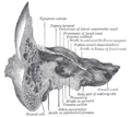 Coronal section of right temporal bone. Prominence of the facial canal labeled at top, fourth from the left.


