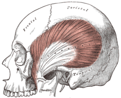
Temporal fascia
| Temporal fascia | |
|---|---|
 | |
 Muscles of the head, face, and neck. (Temporal fascia labeled at top center.) | |
| Details | |
| Identifiers | |
| Latin | fascia temporalis |
| TA98 | A04.1.04.013 |
| TA2 | 2138 |
| FMA | 76863 |
| Anatomical terminology | |
The temporal fascia (or deep temporal fascia[1]: 357 ) is a fascia of the head that covers the temporalis muscle and structures situated superior to the zygomatic arch.[2]
The fascia is attached superiorly at the superior temporal line; inferiorly, it splits into two layers at the superior border of the zygomatic arch - the superficial layer then attaches to the lateral aspect of the superior border of the arch, and the deep layer to its medial aspect.[1]: 357
The space between the two layers is occupied by adipose tissue and contains a branch of the superficial temporal artery, and the zygomaticotemporal nerve.[1]: 357
Anatomy
The temporal fascia t is a strong fibrous investment.[citation needed]
Structure
Superiorly, it is a single layer, attached to the entire extent of the superior temporal line.[citation needed]
Inferiorly, where it is fixed to the zygomatic arch, it consists of two layers, one of which is inserted into the lateral, and the other into the medial border of the arch.[citation needed]
Contents
A small quantity of fat, the orbital branch of the superficial temporal artery, and a filament from the zygomatic branch of the maxillary nerve, are contained between the two layers created by the inferior split of the fascia.[citation needed]
Attachments
The superficial fibers of the temporalis muscle attach onto the deep surface of the temporal fascia.[citation needed]
Relations
Superficial to the temporal fascia are the auricularis anterior muscle and superior auricular muscle, the galea aponeurotica, and (part of) the orbicularis oculi muscle.[citation needed]
The superficial temporal vessels and the auriculotemporal nerve cross it inferoposteriorly.[citation needed]
The parotid fascia proceeds to the temporal fascia.[citation needed]
Text is available under the CC BY-SA 4.0 license; additional terms may apply.
Images, videos and audio are available under their respective licenses.
