For faster navigation, this Iframe is preloading the Wikiwand page for Fil:Spindle centriole - embryonic brain mouse - TEM.jpg.

Fil:Spindle centriole - embryonic brain mouse - TEM.jpg
Fuld opløsning(1.283 × 1.600 billedpunkter, filstørrelse: 901 KB, MIME-type: image/jpeg)


Denne fil er fra Wikimedia Commons

Beskrivelse

Beskrivelse

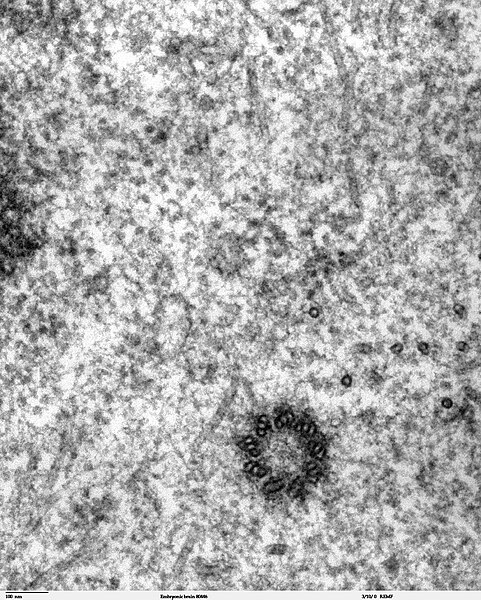
Transmission electron microscope image of a thin section cut through the developing brain tissue (telencephalic hemisphere) of an 11.5 day mouse embryo. This high magnification image of "Embryonic brain 80445" show a spindle centriole and some spindle microtubules visible in the cytoplasm of a mitotic cell at the luminal surface of the telencephalon.

JEOL 100CX TEM

References:

Marin-Padilla, M. (1985) "Early Vascularization of the Embryonic Cerebral Cortex: Golgi and Electron Microscope Studies", J. Comparative Neurology, 241:237-249

Marin-Padilla, M. and M. Amievo (1989) "Early Neurogenesis of the Mouse Olfactory Nerve: Golgi and Electron Microscope Studies", J. Comparative Neurology, 288:339-352
Kilde
Forfatter Louisa Howard, Miguel Marin-Padilla
Tilladelse
(Genbrug af denne fil)
PD

Licensering

Public domain Dette værk er blevet udgivet som offentlig ejendom af dets skaber, Louisa Howard, Miguel Marin-Padilla. Dette gælder i hele verden.
I nogle lande er dette ikke juridisk muligt. I så fald:
Louisa Howard, Miguel Marin-Padilla giver enhver ret til at anvende dette værk til ethvert formål, uden nogen restriktioner, medmindre sådanne restriktioner er påkrævede ved lov.

Captions

Tilføj en kort forklaring på en enkelt linje om hvad filen viser

Elementer som er med i denne fil

afbilder

image/jpeg

Filhistorik

Klik på en dato/tid for at se filen som den så ud på det tidspunkt.

Dato/tidMiniaturebilledeDimensionerBrugerKommentar
nuværende3. nov. 2006, 00:02Miniature af versionen fra 3. nov. 2006, 00:021.283 × 1.600 (901 KB)Patho((Information |Description=Transmission electron microscope image of a thin section cut through the developing brain tissue (telencephalic hemisphere) of an 11.5 day mouse embryo. This high magnification image of "Embryonic brain 80445" show a spindle cen

Filanvendelse

Den følgende side bruger denne fil:

Global filanvendelse

Følgende andre wikier anvender denne fil:

Metadata

{{bottomLinkPreText}} {{bottomLinkText}}
Fil:Spindle centriole - embryonic brain mouse - TEM.jpg
Listen to this article

This browser is not supported by Wikiwand :(
Wikiwand requires a browser with modern capabilities in order to provide you with the best reading experience.
Please download and use one of the following browsers:

This article was just edited, click to reload
This article has been deleted on Wikipedia (Why?)

Back to homepage

Please click Add in the dialog above
Please click Allow in the top-left corner,
then click Install Now in the dialog
Please click Open in the download dialog,
then click Install
Please click the "Downloads" icon in the Safari toolbar, open the first download in the list,
then click Install
{{::$root.activation.text}}

Install Wikiwand

Install on Chrome Install on Firefox
Don't forget to rate us

Tell your friends about Wikiwand!

Gmail Facebook Twitter Link

Enjoying Wikiwand?

Tell your friends and spread the love:
Share on Gmail Share on Facebook Share on Twitter Share on Buffer

Our magic isn't perfect

You can help our automatic cover photo selection by reporting an unsuitable photo.

This photo is visually disturbing This photo is not a good choice

Thank you for helping!


Your input will affect cover photo selection, along with input from other users.

X

Get ready for Wikiwand 2.0 🎉! the new version arrives on September 1st! Don't want to wait?